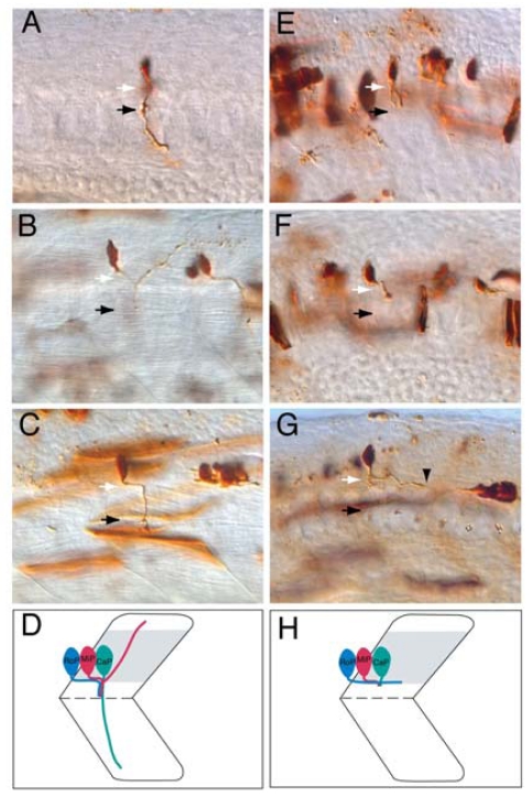
Figure 4.
diwanka is required for motor axon exit in zebrafish embryos. (A–D): Wild-type axonal projections of CaP (A), MiP (B), and RoP (C) primary MNs labeled with fixable dyes as previously described [12]. White arrows indicate the lower half of the spinal cord, which is out of the focal plane, whereas black arrows indicate the choice point, the distal end of a common path followed by CaP, MiP, and RoP motor growth cones prior to their divergence into ventral, dorsal and medial myotomal regions, respectively (D); (E–H): In diwanka mutants, CaP (E) and MiP (F) axons extend wild-type projections within the spinal cord, however they exhibit abnormal projections along the common path (black arrow). Notably, the majority of RoP axons fail to exit the spinal cord in diwanka mutants and instead extend their axons caudally within the CNS (black arrowhead points to a RoP growth cone) (G,H) [12].

