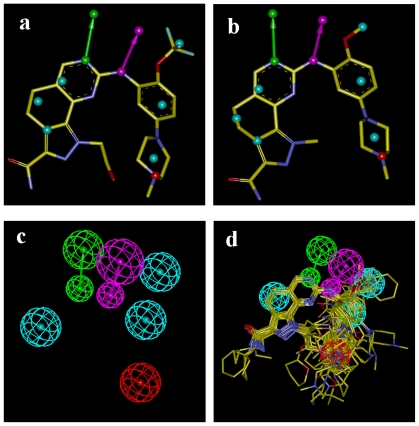
Figure 4.
(a) Pharmacophore model derived from 2YAC; (b) Pharmacophore model derived from 3KB7; (c) The merged model; (d) The compounds alignment based on the merged model. Features are color-coded with magenta for hydrogen-bond donor, green for hydrogen-bond acceptor, light-blue for hydrophobic, red for ionizable positive.

