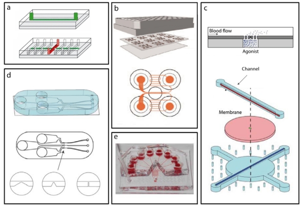
Figure 3.
Microfluidic Devices: (a) A microfluidic device with seven parallel microchannels (red) to run simultaneous experiments under different shear rates. A horizontal channel (green) is used to flow ECM proteins in solution and deposit the protein as adhesive patches on the array of horizontal channels used for blood flow. Adapted with permissions from [77]. (b) A compact array of microfluidic devices allows the operator to run multiple experiments simultaneously. Adapted with permissions from [82]. (c) A microfluidic device used for the controlled release of agonist through a membrane structure at the bottom of the flow channel. Adapted with permissions from [73]. (d) A microfluidic device with three different types of obstructive geometries in the main channel is used to examine the effect of gradients in the strain rate. Adapted with permissions from [75]. (e) A microfluidic device used to expose the same sample of blood to different inhibitors. Adapted with permissions from [83].

