Figure 3.
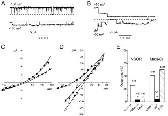
Two types of microscopic currents activated in osmotically swollen thymocytes. (A) Single VSOR anion channel activity. (B) Single maxi-anion channel activity. Pipette: 100 mM TEACl-pipette solution; bath: hypotonic high-K+ solution. Dashed lines correspond to zero current level. (C) I-V relationship for the single-channel events similar to those shown in (A). Unitary currents were recorded in the cell-attached mode with standard 100 mM CsCl-pipette solution (open circles), 100 mM TEACl-pipette solution (closed diamonds) and 30 mM CsCl-pipette solution (filled triangles). Each data point represents the mean ± SEM of 5 to 29 measurements from 8–14 different patches. (D) I-V relationship for the single-channel events similar to those shown in (B). Unitary currents were recorded in the cell-attached mode with 100 mM TEACl-pipette solution and hypotonic high-K+ solution in the bath (filled triangles), or in inside-out mode with Ringer solution in the pipette and in the bath (open circles). Filled diamonds and filled squares: 135 mM NaCl in the bath solution was replaced with equimolar TEACl and Na-glutamate, respectively (inside-out, pipette filled with Ringer solution). Each data point represents the mean ± SEM of 5 to 30 measurements from 7–10 different patches. (E) Effects of phloretin (200 μM), DIOA (10 μM) and Gd3+ ions (50 μM) on the occurrence of the VSOR-type and maxi-anion-type channels in the on-cell mode (the drugs added to the pipette solution). On the top of the bars: number of channel-containing patches/total number of patches. * Significantly different from the control value at P < 0.05.

