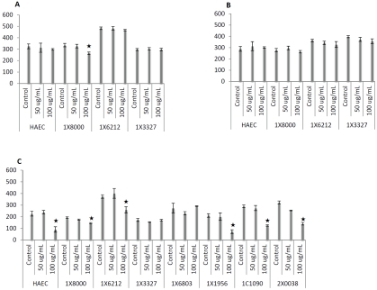
Figure 2.
Effects of three LDL preparations at 50 or 100 μg/mL on endothelial cell growth for 24 hours. Total protein content of lysates was used as an indicator of cell growth, using human aortic endothelial cells (HAEC) as a reference. LDL products at indicated dosages were added to endothelial cell cultures (as indicated by animal ID). Panel A shows that native LDL at 50 μg/mL did not affect growth of cells from any of the four baboons, but a significant decrease in total protein was observed in one culture (1×8000) at 100 μg/mL. Panel B shows that no significant differences in inhibition of cell growth were observed between control cultures and those incubated with minimally oxidized LDL at either concentration. Panel C shows five of seven subjects exhibited significant cell death after exposure to 100 μg/ mL oxidized LDL by comparison with their corresponding controls. * indicates p<0.05.

