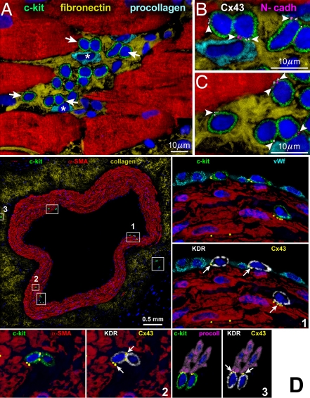
Figure 1.
Niches in the normal human heart. (A-C) Cluster of c-kitPOS cells (green). Arrows in A define the areas in B and C. Gap (connexin 43: Cx43, white; arrowheads) and adherens (N-cadherin: N-cadh, magenta; arrowheads) junctions are shown at higher magnification. Cx43 and N-cadh are present between c-kitPOS cells and myocytes (α-SA, red) and fibroblasts (procollagen, light blue) [1]. (D) Cross-section of human epicardial coronary artery composed of several layers of smooth muscle cells (α-smooth-muscle-actin: α-SMA, red). The c-kit-positive cells (green) are included in six rectangles. Three of the six rectangles are shown at higher magnification in the adjacent panels. The c-kit-positive cells express KDR (white). Cx43 (yellow dots; arrows) is seen between c-kit-KDR-positive cells and endothelial cells (von Willebrand factor: vWf, bright blue), smooth muscle cells (α-SMA, red), and adventitial fibroblasts (procoll, magenta) [2].

