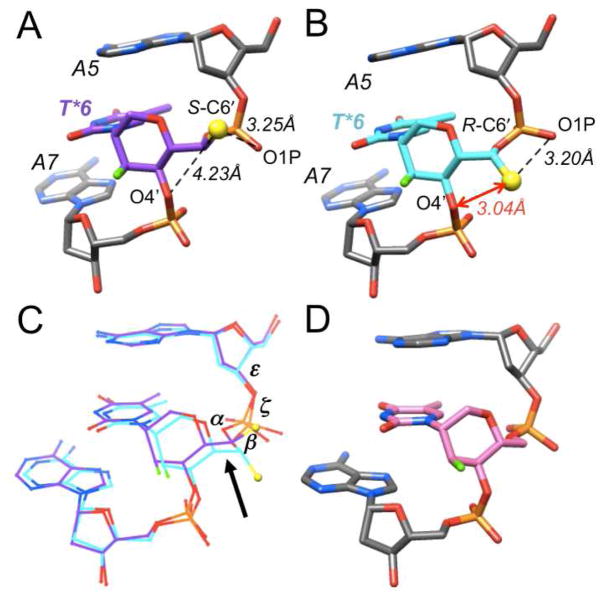
Figure 2.
Conformations of (A) S- and (B) R-6′-Me-FHNA (purple and cyan carbon atoms, resp.), (C) superimposition of the two, and (D) the conformation of FHNA (pink carbon atoms) for comparison. The methyl carbon is shown as a yellow sphere, F3′ is green, residues are labeled and the short 1···5 contact in R-6′-Me-FHNA T is highlighted with a red arrow.

