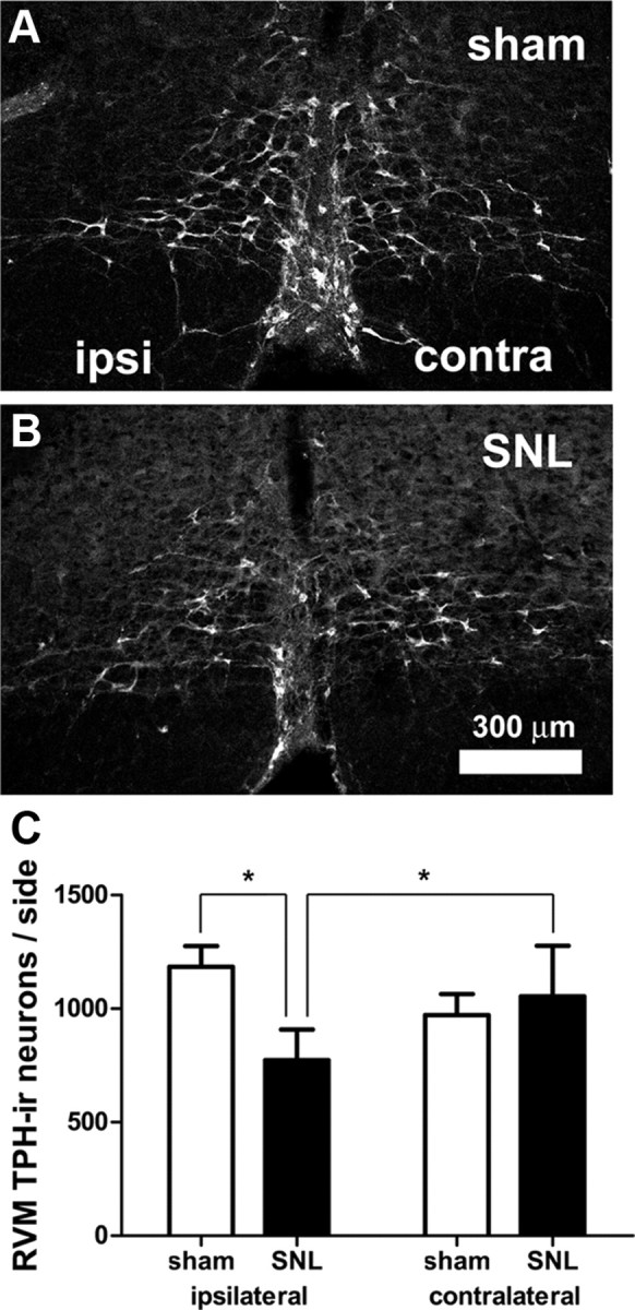
Figure 3.

SNL loss of TPH-IR neurons in the RVM. A, B, Confocal images showing TPH-IR in the RVM in sham-operated (A) and SNL-treated (B) rats. Parameters for image acquisition and display (brightness, contrast, filtering) were identical. “Ipsilateral” and “contralateral” are relative to the nerve ligation. C, The number of RVM TPH-IR neurons ipsilateral to SNL was significantly less than contralateral to SNL. In addition, the number was significantly less than that found ipsilateral to sham-surgery (*p < 0.05; two-way ANOVA). TPH-IR neurons were counted based on their Nissl counterstaining, as described in Materials and Methods. Scale bar in B applies to A and B.
