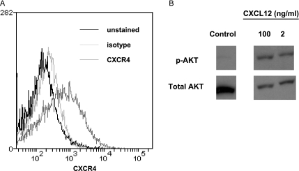
Figure 1.
HeyA8 ovarian cancer cells express functional CXCR4. (A) Cell surface levels of CXCR4 on HeyA8 cells were determined by flow cytometry. Dark line indicates unstained; light line, isotype antibody control; and intermediate line, CXCR4 antibody. (B) HeyA8 cells were serum starved overnight and then incubated with 2 or 100 ng/ml CXCL12 for 10 minutes. Cell lysates were probed for phosphorylated AKT (p-AKT, active form) and total AKT as a control for protein loading.

