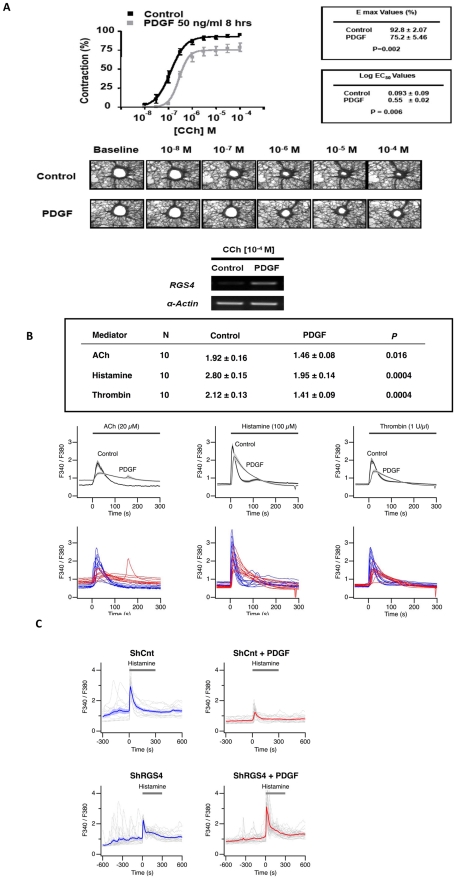
Figure 4. PDGF markedly inhibits carbachol-induced bronchoconstriction in human small airways.
(A) PCLS were obtained from healthy donors and treated with medium or PDGF (50 ng/ml) for 8 h followed by analysis of carbachol-induced small airway constriction by microscopy. Log EC50 and Emax values (mean ± SEM) were determined in experiments on 16 airways obtained from 4 separate donors as described in Methods. (B) PDGF attenuates agonist-induced increases in [Ca2+]i. HASM cells were stimulated for 8 h with 10 ng/ml PDGF or diluent followed by measurement of [Ca2+]i using a Ca2+-sensing fluorophore after stimulation with acetylcholine (ACh), histamine or thrombin. Single-cell calcium transients were measured over a period of 300 sec. Table shows mean ± SEM of peak [Ca2+]i levels determined in 30 cells (P values determined by 2-tailed Student's t test). Bottom tracings represent [Ca2+]i in individual cells (blue line = control, diluent-treated; red line = PDGF-treated). Middle tracings represent group mean data ± SEM shown in the shaded segments. (C) RGS4 is required for PDGF-mediated attenuation of agonist-induced [Ca2+]i in HASM cells. The mean responses are shown using thick curves, and the individual cell responses are shown using dashed curves.

