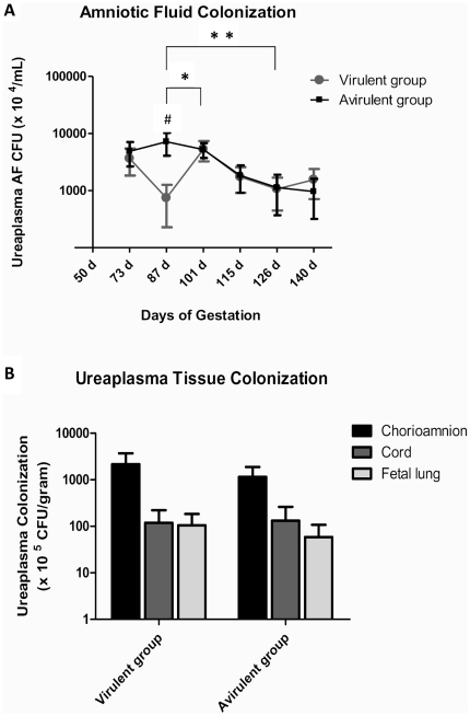
Figure 1. Ureaplasma colonization of amniotic fluid and fetal tissues.
(A) Chronic infection of the amniotic fluid was observed in all ewes experimentally infected with ureaplasmas from the time of inoculation (55 d) until fetuses were delivered at 140 d. Amniotic fluid ureaplasma colonization was significantly increased in the avirulent group when compared to the virulent group at 87 d (p<0.05, denoted by #). Statistically significant differences in amniotic fluid ureaplasma colonization within animal groups occurred between 87 d and 101 d; and 87 d and 126 d (p<0.05). (B) Ureaplasmas were isolated from the chorioamnion, cord and fetal lung; however, recovered ureaplasma CFU/g was not different between animal groups for the tested tissue types. * = statistically significant difference between time points in the virulent group only; ** = statistically significant difference between time points in both groups. AF = amniotic fluid; CFU = colony forming units; d = days of gestation. Data are presented as mean ± SEM.

