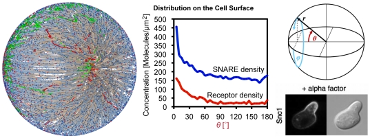
Figure 9. Cell Polarization.
Molecules, vesicle paths and cytoskeleton structure. SNAREs (blue) and Receptors (red) accumulate on the left. Endocytic vesicle tracks are shown in red, recycling paths in green. The polarization of the cell is in agreement with the findings of Valdez-Taubas and Pelham [58] for the SNARE Snc1. The microscope image is reprinted from (Valdez-Taubas and Pelham, 2003) with permission from Elsevier.

