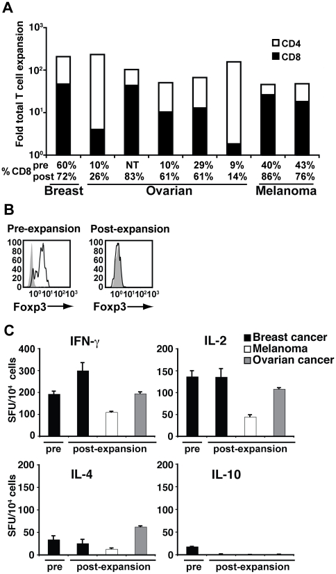
Figure 4. aAPC/mOKT3 expanded TIL are Foxp3 negative and secrete predominantly Th1 cytokines.
(A) Expansion of TIL obtained from breast and ovarian cancer ascites and melanoma metastases is shown. Shading indicates the proportion of CD4+ (white) and CD8+ (black) T cells in expanded cultures. The percentage of CD8+ T cells in pre- and post-expansion cultures is shown. Note that in all samples tested, the percentage of CD8+ T cells increased even in those that initially contained a minimal percentage of CD8+ T cells. NT denotes not tested. (B) CD4+ CD25+ Foxp3+ Treg cells, present pre-expansion, were not detectable after one month of culture. CD4+ CD25+ cells were intracellularly stained with anti-Foxp3 mAb (open) and isotype control (shaded). (C) IFN-γ, IL-2, IL-4, and IL-10 secretion of expanded TIL was determined by ELISPOT assays. Cytokine secretion by TIL from the breast cancer ascites specimen prior to expansion is shown as a control. Pre-expansion samples from melanoma and ovarian cancer specimens were not studied because of low initial cell numbers.

