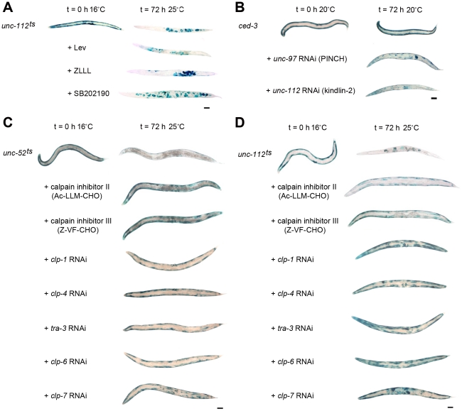
Figure 7. Calpain inhibition blocks protein degradation induced by acute loss of attachment.
A) Age synchronised L1 stage unc-112ts mutants were gown to young adulthood at 16°C (t = 0 h) before being temperature shifted to 25°C and cultured for a further 72 h under normal conditions (top row), or on plates seeded with bacteria plus proteasome pathway inhibitors (+Lev, second row; +ZLLL, third row) or a lysosome pathway inhibitor (+SB202190, fourth row). B) ced-3 (caspase 3) mutants were age synchronised at the L1 stage and grown to adulthood at 20°C (t = 0 h) before being cultured on RNAi plates against unc-97 or unc-112 for a further 72 h. In both A and B, at t = 0 h and 72 h approximately 20–30 animals were stained for β-galactosidase activity (blue). C) unc-52ts and D) unc-112ts mutants were cultured for two generations at 16°C (permissive, mutation inactive) under either normal, control conditions or on RNAi against clp-1, clp-4, tra-3, clp-6 or clp-7. Second generation animals were then age synchronised at L1 stage and grown to young adulthood at 16°C (t = 0 h) before being temperature shifted to 25°C (non-permissive, mutation active) and cultured on the respective condition for a further 72 h. Some temperature shifted animals were also placed on calpain inhibitor II or calpain inhibitor III drug plates (5 µg/ml) at t = 0 h and cultured on drug plates for a further 72 h. Approximately 20–30 animals were stained for β-galactosidase (blue) at t = 0 h, and at 24 h, 48 h (not shown) and 72 h. All experiments were performed at least three times. The blue stain that appears as circles in the centre of t = 72 h animals is stain in muscles of developing embryos (for example unc-112ts in A and D). Scale bars represent 100 µm.

