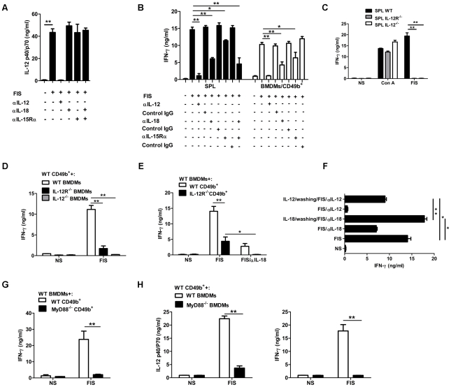
Figure 2. Network of cytokine dependence of NK cell activation by B. anthracis spores.
Effect of neutralization of IL-12, IL-18 or IL-15Rα on (A) IL-12p40/p70 concentration in culture supernatants of FIS-stimulated BMDMs or (B) IFN-γ production by splenocytes (SPL; left panel), or purified CD49b+ cells co-cultured with BMDMs (right panel). (C) Splenocytes (SPL) from wild-type (WT), IL-12R−/− or IL-12−/− C57BL/6 mice were stimulated with FIS, or ConA as a positive control. (D) CD49b+ cells from WT C57BL/6 mice were co-cultured with BMDMs from IL-12R−/− or IL-12−/− C57BL/6 mice in the presence of FIS. (E) CD49b+ cells from WT or IL-12R−/− C57BL/6 mice were co-cultured with BMDMs from WT C57BL/6 mice in the presence of FIS with or without IL-18 neutralizing antibody. (F) Effect of short-term priming with IL-12 or IL-18 on spore-stimulation of splenocytes; corresponding cytokine neutralization was maintained for the remainder of the assay. (G) Purified CD49b+ cells from WT or MyD88−/− C57BL/6 mice were co-cultured with BMDMs from WT C57BL/6 in the presence of FIS. (H) Purified CD49b+ cells from C57BL/6 WT mice were co-cultured with BMDMs from WT or MyD88−/− C57BL/6 mice in the presence of FIS; IL-12 (left panel), or IFN-γ (right panel) production. For all experiments with purified CD49b+ cells, no IFN-γ was detected after direct stimulation with spores (D, E, G, H; data not shown). For all experiments, values are mean ± SD for at least three measurements and are representative of at least three independent experiments. Significant differences between experimental conditions are indicated with asterisks (t test; *, P<0.05; **, P<0.01).

