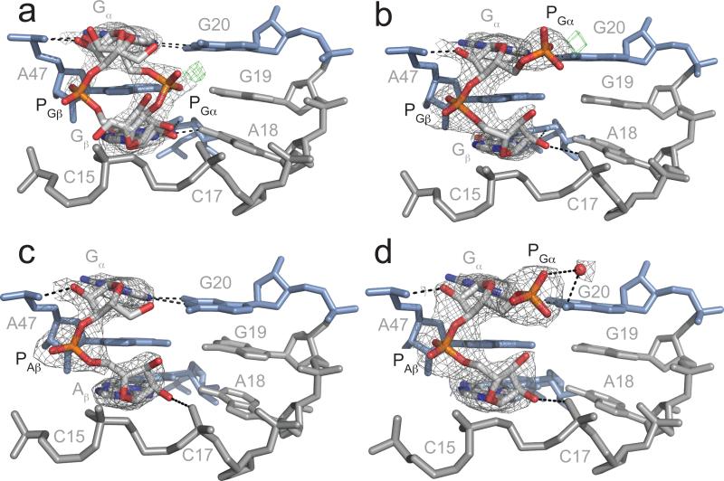
Figure 3.
Structures of linear analogs bound to the c-di-GMP-I riboswitch. Coloring is the same as in Figure 1. 2Fo-Fc density is shown in gray, contoured to 1σ. Fo-Fc density is shown in green. a. Complex with GpG. Both orientations of the ligand are shown. Fo- Fc density is contoured to 3.0σ. b. Complex with pGpG. Fo-Fc density is contoured to 2.4σ. c. Complex of GpA with the C92 mutant riboswitch. d. Complex of pGpA with the C92U mutant riboswitch. The water molecule is shown as a red sphere.

