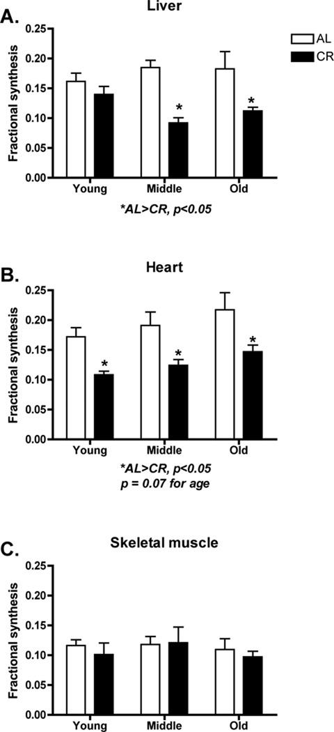
Figure 2.
Cellular proliferation as measured by DNA synthesis over a 6-week period in young, middle, and old AL or CR mice. In liver (A) and heart (B) there were significant decreases in cellular proliferation in CR mice. In heart, there was a trend (p = 0.07) for increased cellular proliferation with age. In skeletal muscle there were no differences in cellular proliferation (C) although it is worth noting that there was measureable DNA synthesis in skeletal muscle. n = 6–8 per group.

