
Fig. 4.
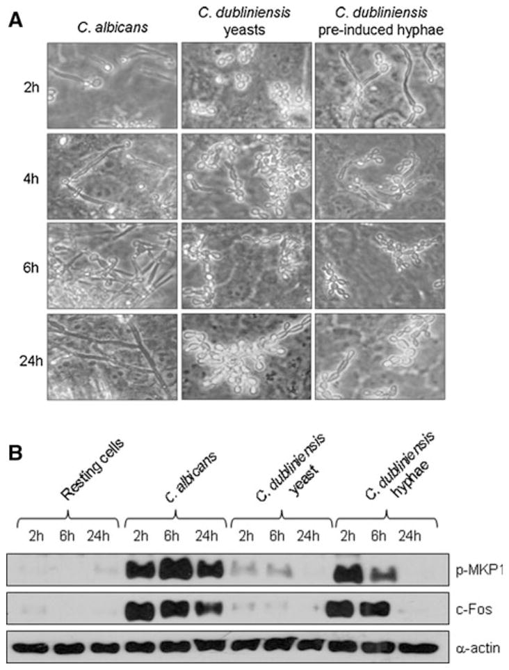
Reduction of MKP1 and c-Fos activation upon reversion of C. dubliniensis hyphae to pseudohyphal/yeast cells. Candida dubliniensis was pre-induced to form hyphae in water/10% serum for 3 h at 37°C and added to TR146 cells. At 2, 6 and 24 h postinfection a the morphology of C. dubliniensis or b MKP1 phosphorylation and c-Fos production was assessed by microscopy or Western blot, respectively. Bands are shown relative to α-actin loading control. A fungal/epithelial cell MOI of 10:1 was used for the 2 and 6 h time points and 0.01 for the 24 h time point. Data are representative of three independent experiments