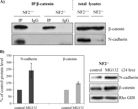
Figure 3.
N-cadherin reduced in schwannoma cells and its degradation. (A) Schwann (NF2+/+) and schwannoma cells (NF2-/-) were lysed in NP40 buffer and immunoprecipitated with an antibody against β-catenin. IgG mouse without anti-β-catenin antibody served as control. Western blot analysis was carried out to detect the levels of β-catenin and N-cadherin in both co-IP complex and total lysates. (B) N-cadherin is accumulated after MG132 inhibition. Schwannoma cells were treated with MG132 (1 µM) for 24 hours, Western blots were then carried out for N-cadherin and β-catenin. RhoGDI served as loading control. Error bars represent the mean ± SEM.

