Figure 1.
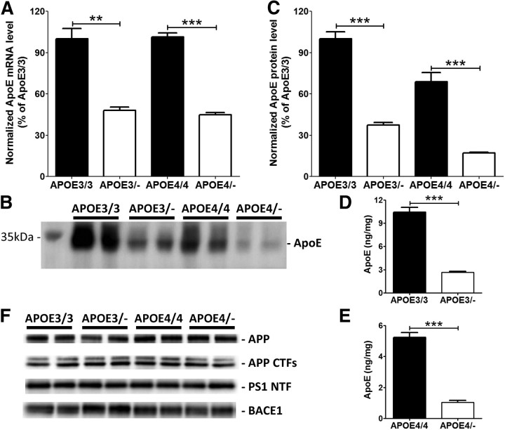
Reduction of apoE levels in human APOE haploinsufficient mice. Cortex from APOE homozygous (APOE3/3 and APOE4/4) and hemizygous (APOE3/− and APOE4/−) mice were used to measure apoE mRNA and protein levels. A, ApoE mRNA levels were measured by quantitative real-time PCR. B, Levels of PBS-soluble apoE were assessed by probing a membrane with anti-apoE antibody. C, Quantitative analyses of Western blots were performed with tubulin normalization (n = 4 per genotype). D, E, To validate the Western blot data, apoE levels were also measured by using an apoE-specific ELISA from APOE3/3 and APOE3/− mice (D) and APOE4/4 and APOE4/− mice (E) (n = 9–18 per genotype). F, Levels of APP, APP CTFs, PS1 NTF, and BACE1 proteins were measured by Western blotting (n = 4 per genotype). Levels of these proteins after normalizing with tubulin signals were not significantly altered by APOE haploinsufficiency. All graphs represent values in mean ± SEM.

