
Figure 3.
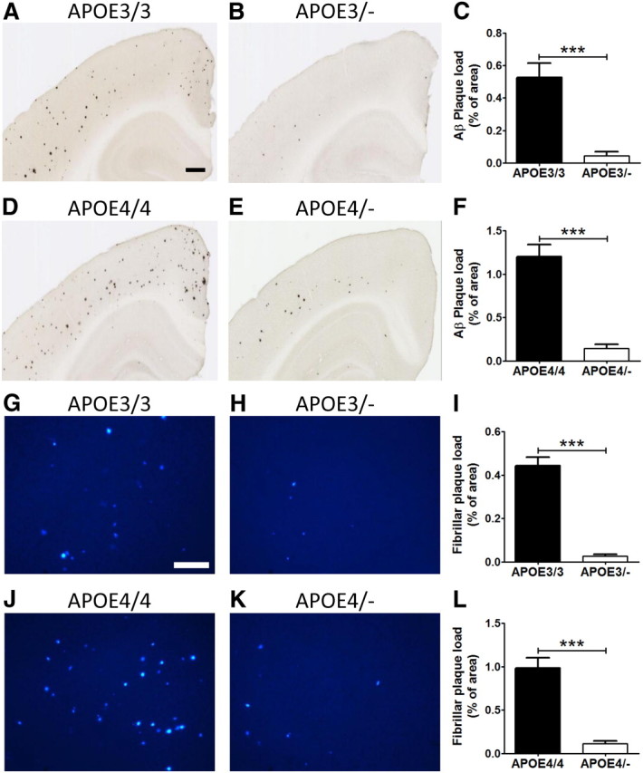
Haploinsufficiency of human APOE leads to reduction of Aβ and fibrillar Aβ plaque deposition. A, B, D, E, Brain sections from 3-month-old APOE homozygous (A, D) and hemizygous (B, E) mice were immunostained for amyloid with anti-Aβ antibody (HJ3.4-biotin). Scale bars, 400 μm. A–C, The extent of Aβ deposition detected by HJ3.4-biotin antibody was quantified (C) from cortex of APOE3/3 (A) and APOE3/− mice (B). D–F, The extent of Aβ deposition detected was quantified (F) from cortex of APOE4/4 (D) and APOE4/− mice (E). G, H, J, K, Brain sections from 3-month-old APOE homozygous (G, J) and hemizygous (H, K) mice were stained with X-34 dye that recognizes only fibrillar plaques. Scale bar, 200 μm. G–I, Fibrillar plaque load detected by X-34 dye was quantified (I) from cortex of APOE3/3 homozygous (G) and APOE3/− mice (H). J–L, The extent of fibrillar plaque load was also analyzed (L) from cortex of APOE4/4 (J) and APOE4/− mice (E). (n = 12–20 per genotype).