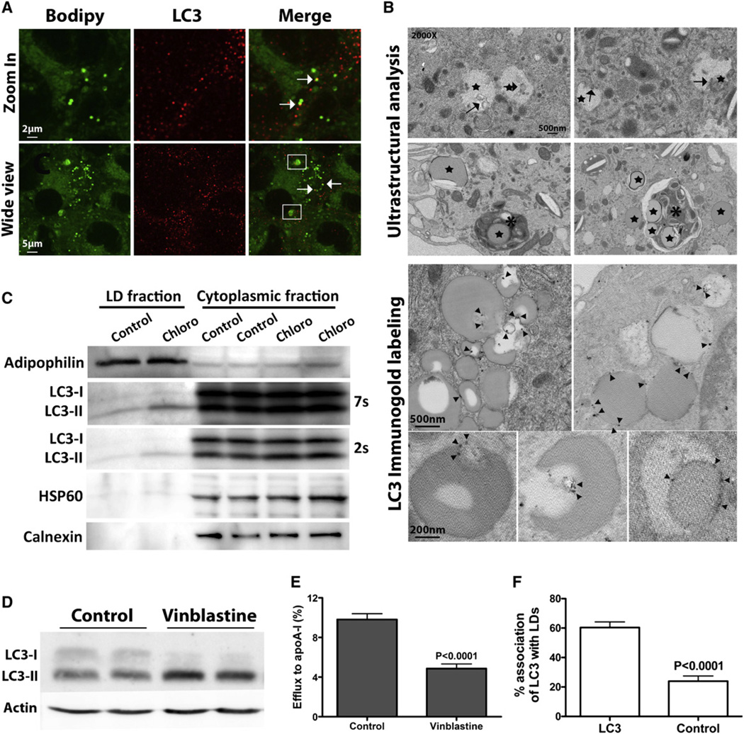
Figure 2. Autophagy Is Implicated in Cytoplasmic LD Degradation.
(A–C) In lipid-loaded BMDMs, direct association of autophagomes with LDs is observed by immunofluorescence (A), electron microscopy (B), and in an isolated LD fraction (C).
(D) Vinblastine treatment inhibits autophagosome degradation, as shown by elevated LC3-II.
(E) Inhibition of autophagy by vinblastine treatment during cholesterol efflux decreases efflux to apoA-I.
(F) Quantification of LDs containing gold particles (cells immunostained with LC3 are compared to the secondary antibody alone negative control).

