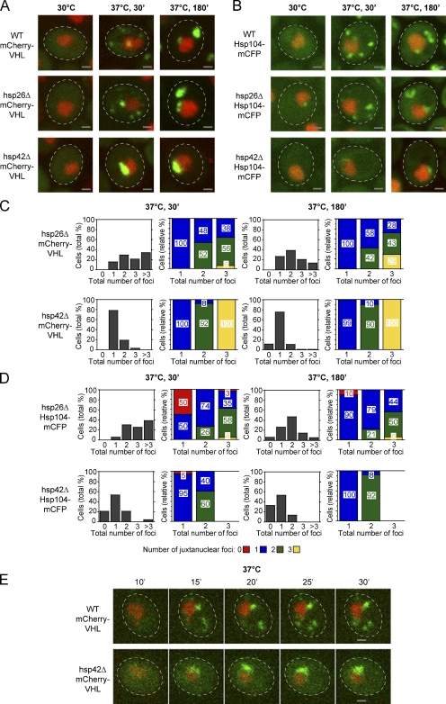
Figure 2.
Hsp42 is essential for the targeting of misfolded proteins to peripheral aggregates. (A and B) Time-dependent changes in the localization of mCherry-VHL (A) and Hsp104-mCFP (B; both green) at 30°C and after a shift to 37°C for 30 and 180 min in the isogenic WT, hsp26Δ, and hsp42Δ strains. MG132 was added before the temperature shift. Nuclei were visualized by coexpressing HTB1-cerulean or HTB1-mCherry (red). (C and D) Number (shaded bars) and localization (colored bars) of mCherry-VHL (C) and Hsp104-mCFP (D) aggregates in hsp26Δ and hsp42Δ cells after incubation at 37°C for 30 and 180 min. MG132 was added before the temperature shift. The total number of foci per cell is depicted on the x axis in all diagrams. Quantifications are based on the analysis of 70–90 cells and are representatives of at least two experimental repeats. (E) In hsp42Δ cells, mCherry-VHL foci form exclusively at the nucleus. Time-lapse microscopy pictures of single WT and hsp42Δ cells expressing mCherry-VHL (green) after a shift to 37°C for the indicated time period. MG132 was added before the temperature shift. Nuclei were visualized by coexpressing HTB1-cerulean (red). The dashed lines indicate the border of respective yeast cells. Bars, 1 µm.

