Figure 2.
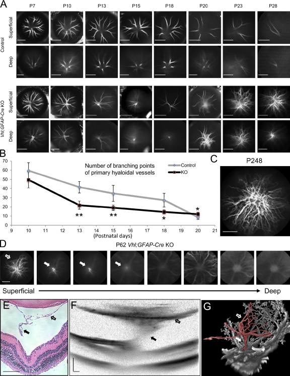
Development of persistent hyaloidal vasculature in Vhl;GFAP-Cre knockout (KO) mice. (A–C) The conditional deletion of Vhl in astrocytes using GFAP-Cre induces early accelerated regression of hyaloidal vessels followed by massive secondary outgrowth. (A) Representative in vivo images of hyaloidal vasculature in two different dimensional depth of Vhl mutants (third and fourth rows) and control littermates (first and second rows) from P7 to 28. (B) Quantification of the primary (but not secondary outgrowth of) hyaloidal vasculature at P10, 13, 15, 18, and 20 on a scatter plot (n = 4–6). *, P < 0.05; **, P < 0.01. Error bars indicate mean ± SD. (C) Secondary outgrowth of hyaloidal vasculature observed at stage P248 (∼8 mo). Note that vessels form a stalk formation in the deep optical vitreal regions in early stages (fourth row in A). Significantly fewer branching point numbers of the primary hyaloidal vasculature were observed in Vhl mutants compared with controls during P13 to 18 stages (B). Pronounced secondary outgrowth from the stalk occurs from P20 and persists throughout the lives of the Vhl mutants (A and C). (D–G) Stalk formation observed in CSLO (D), paraffin-embedded section (E), OCT (F), and in vascular casts (G). Note the connecting stalk (closed arrows) between the secondary outgrowth (open arrows) and the optic artery. Bars: (A, C, and D) 2,000 µm; (E and F) 200 µm.

