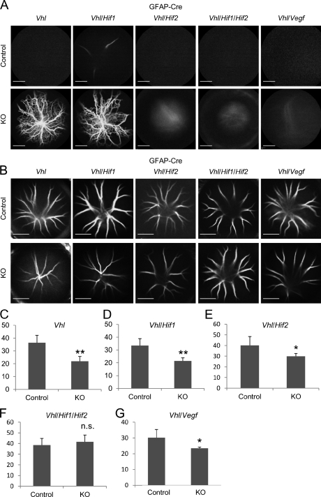
Figure 3.
Secondary outgrowth but not accelerated hyaloidal regression in Vhl mutants is caused via the HIF-2/VEGF signaling pathway. (A) Secondary outgrowth in various combinations of astrocyte-specific knockout (KO) mice at P40. Note that massive secondary outgrowth is observed in Vhl (both HIF-1α and HIF-2α are stabilized) and Vhl/Hif-1α double mutants (only HIF-2α is stabilized). The outgrowth phenotype is prevented in Vhl/Hif-2α (only HIF-1α is stabilized), in Vhl/Hif-1α/Hif-2α, or in Vhl/Vegf mutants, demonstrating that HIF-2α/VEGF signaling is required for the development of secondary outgrowth. (B–G) Representative hyaloidal vascular images (B) and numbers of branching points (C–G) in various combinations of knockout mice at P14 (n = 4–8). Note that accelerated hyaloidal regression is observed in Vhl (C), Vhl/Hif-1α (D), VHL/Hif-2α (E), and Vhl/Vegf (G) but not Vhl/Hif-1α/Hif-2α (F) mutants, demonstrating that HIF-1α and HIF-2α compensate for this phenotype independently from VEGF. n.s., not significant. *, P < 0.05; **, P < 0.01. Error bars indicate mean ± SD. Bars, 2,000 µm.

