Figure 6.
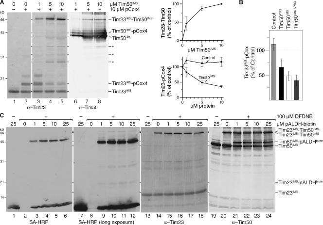
Presequence and Tim50 binding to Tim23 are mutually exclusive. (A) 1 µM purified Tim23IMS was incubated with excess pCox4 in the absence or presence of the indicated amounts of Tim50IMS and subjected to chemical cross-linking by 100 µM DFDNB for 30 min. Samples were analyzed by Western blotting using the indicated antibodies (left). Quantification of Tim23–Tim50 adduct. 100%, adduct formed with maximal amount of Tim50 (SEM; n = 5; top right). Quantification of the Tim23–pCox4 adduct in response to increasing amounts of Tim50IMS or Tim21IMS (control). 100%, adduct formed without additional protein (SEM; n = 5; bottom right). Asterisks denote degradation products of Tim50IMS; Asterisk, Coomassie-stained degradation product of Tim50IMS detected as bleed-through using a fluorescence scanner at 685 nm. (B) 1 µM purified Tim23IMS was incubated with 10 µM pCox4 in the presence of 5 µM of indicated Tim50 constructs or Tim21IMS (control) and subjected to chemical cross-linking. The Tim23IMS–pCox4 adduct was quantified. 100%, adduct formed in the absence of additional protein (SEM; n = 5 for control and Tim50IMS, n = 3 for Tim50PBD, and n = 7 for Tim50ΔPBD). (C) Equimolar amounts (1 µM) of Tim50IMS and Tim23IMS were incubated with the indicated amounts of biotin-labeled pALDH. After chemical cross-linking using 100 µM DFDNB for 30 min the samples were analyzed by SDS-PAGE and Western blotting with the indicated antibodies.

