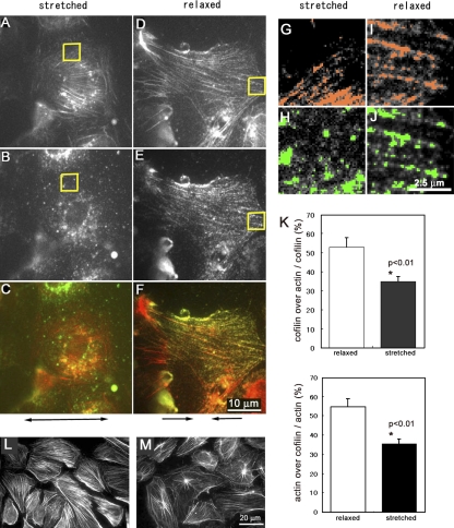
Figure 3.
Cofilin binding to stress fibers is modulated by stretching of semi-intact cells. (A–F) Cofilin did not bind to stress fibers in semi-intact cells that were 10% stretched (A, B, and C) but did bind to stress fibers in the cells when they were 20% relaxed (D, E, and F). The specimen was fixed and then stained for actin (A and D) and cofilin (B and E). These images are merged in C (A and B) and F (D and E). The double-headed arrow below C shows the direction of stretching, and arrows below F show the direction of relaxation. The high magnification images of the areas enclosed by yellow squares in A–D are shown in G–J, respectively. (G–J) Colocalization of actin (colored red in G and I) and cofilin (colored green in H and J) was evaluated by MetaMorph software (measure colocalization). (K) The percentages of cofilin-positive pixels on actin stress fibers (top) and percentages of actin-positive pixels on cofilin-positive pixels (bottom) are shown. Data were obtained from five cells from two independent experiments. Vertical bars denote SEM. (L and M) Stress fibers in semi-intact cells bathed in ATP-free buffer with 250 nM cofilin were not disassembled when cells were kept stretched 10% (L) but were disassembled in the same solution when they were relaxed 20% (M).

