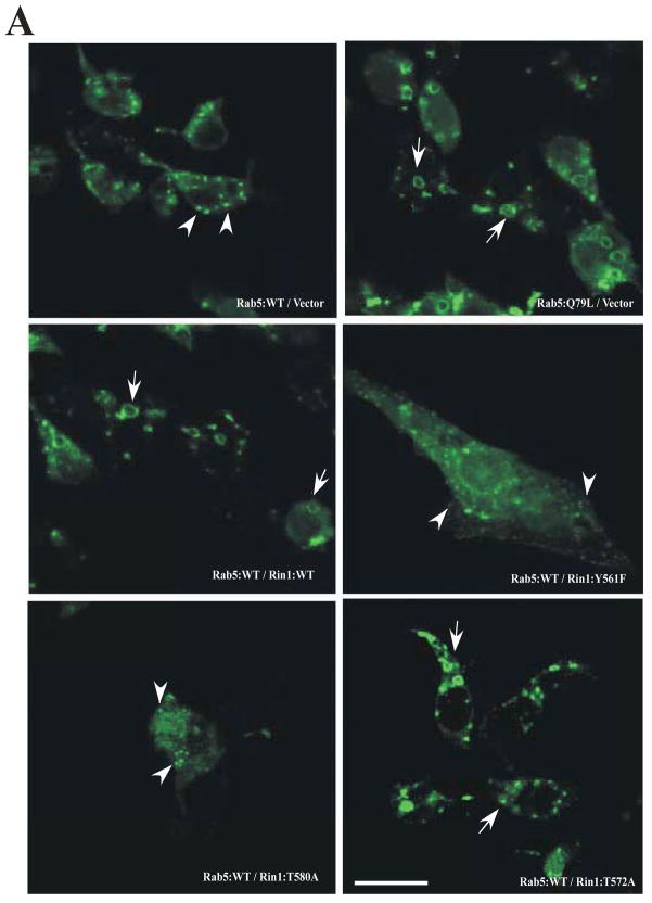
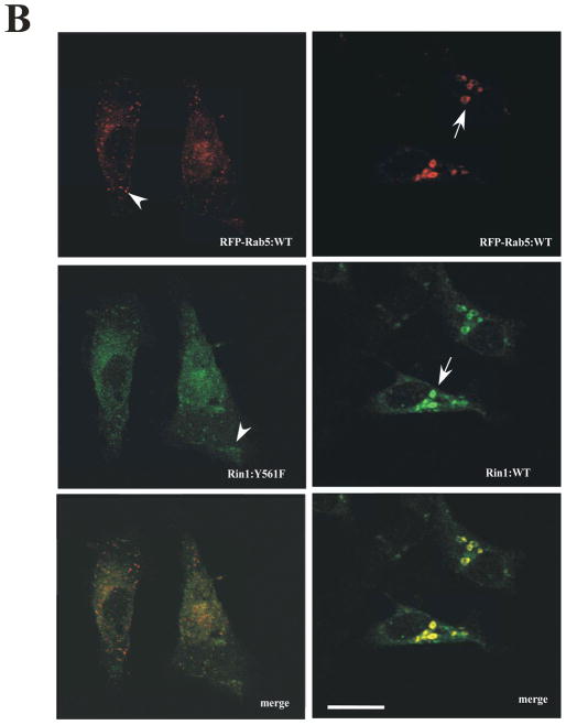
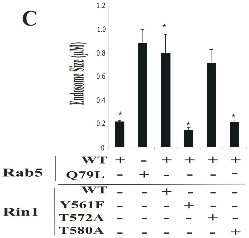
Figure 5. Rin1 mutants affect the formation of enlarged Rab5-positive endosomes.
(A) Stable NR6 cell lines over-expressing either vector alone (control), Rin1: wild type, or Rin1 mutants were transiently transfected with GFP-Rab5: wild type and GFP-Rab5: Q79L mutant. After transfection, cells were processed for immunofluorescence as described in Material and Methods. Confocal fluorescence microscopy images show the morphological changes of Rab5-labeled endosom es in NR6 cells coexpressing both Rin1 and Rab5 constructs as indicated in the Figure. This experiment was repeated three times with similar results. Bar: 10 μm. (B) Stable NR6 cell lines over-expressing either Rin1: wild type or Rin1: Y561F mutant were transiently transfected with RFP-Rab5: wild type. After transfection, the cells were fixed and processed as described in Material and Methods. This experiment was repeated three times with similar results. Bar: 10 μm. The arrowheads and arrows denote the small and enlarged Rab5-positve endosomes, respectively. (C) Different sizes of Rab5-positive endosomes were quantified in control cells (i.e., cells co-expressing Rab5 constructs and vector alone) and cells co-expressing both Rin1 and Rab5 constructs. A total of 2000 Rab5-positive endosomes from 7 cells were used to determine endosome sizes in each experiment. The data are presented as means ± SD of three independent experiments, n=3 *P < 0.001.


