Fig. 1.
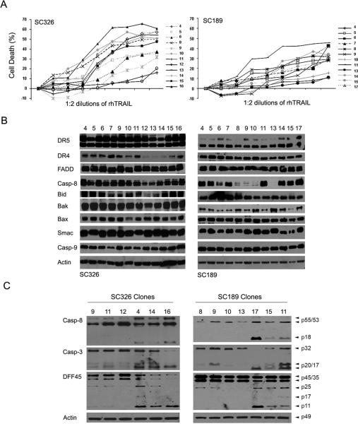
TRAIL-induced apoptosis in non-stem cell clones derived from SC326 and SC189 primary culture. A Each of the clones (indicated to the right) was treated with 1:2 serial dilutions of rhTRAIL starting at 300 ng/ml for 24 hr and the percentage of cell death was assessed by a luminescent cell viability assay. Data are mean +/− SEM (n = 6). B Western blot analysis of the expression of TRAIL apoptotic proteins (indicated to the left) in SC326 and SC189-derived non-stem cell clones. Actin was used as protein-loading control. C Western blot detection of cleavage of caspase-8 (Casp-8), caspase-3 (Casp3) and DFF45 (indicated to the left) in SC326 and SC189 clones after treatment with 300 ng/ml TRAIL for 6 hr.

