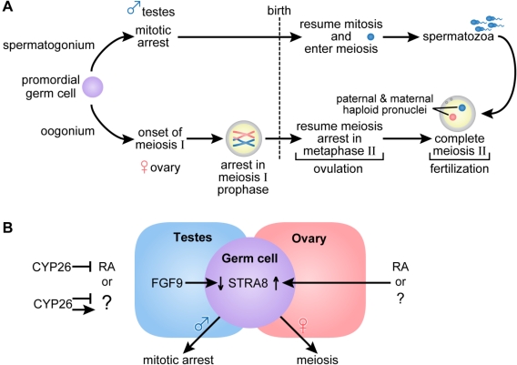
Figure 3.
(A) Germ cell development and gametogenesis. Primordial germ cells colonize the gonad in both male and female embryos. The first morphological marker of sex-specific germ cell development is seen in the female embryo when the oogonia enter meiosis. Primary oocytes proceed through the leptotene, zygotene and pachytene stages of meiotic prophase before birth, when they arrest in diplotene of meiosis I. At ovulation, meiosis I is completed, and the secondary oocytes enter meiosis II and arrest again in metaphase. Meiosis II is completed after fertilization. In the male embryo, germ cells are committed to the spermatogenic program but arrest in G0/G1, and do not complete mitosis and enter meiosis until after they are born. Primary spermatocytes entering meiosis I are seen during the first week of life; secondary spermatocytes complete meiosis II forming spermatids and functional gametes called spermatozoa or sperm. In the male, waves of meiosis continue throughout life. (B) In the female embryo access to RA or alternatively, another factor indicated as (?), promotes entry into meiosis whereas embryonic male germ cells are maintained in a pluripotent state. Either RA or another factor acts in the embryonic female germ cell to increase Stra8, essential for entry into meiosis. In the ovary, Fgf9 levels are low. In the male embryo, entry into meiosis is prevented by the action of CYP26B1; high levels of Fgf9 in the testes antagonize Stra8 expression and maintain germ cells in a pluripotent state. (Adapted from [77,78]).

