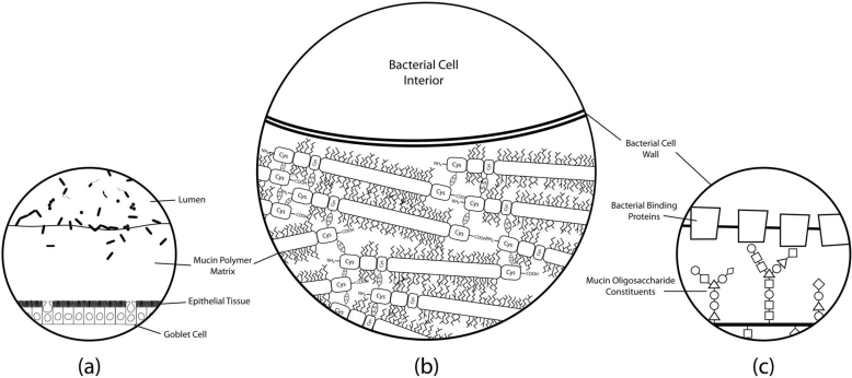
Figure 2.
Simplified histological cross-section of microbial adhesion to the colonic mucosal surface at various magnifications. (a) The layer of mucus atop colonic epithelial villi. Goblet cells can be seen interspersed throughout the columnar enterocytes, producing secretory mucin that makes up the gel matrix. The microbial communities residing in and on top of the mucus layer can only be found at substantial concentrations in the outermost regions of the mucus; (b) The mucus-bacteria interface. The mucin molecules polymerize to form the mucus layer matrix to which cells adhere. Extensive disulfide bonding between cysteine-rich regions of the mucin protein cores creates the characteristic viscoelastic properties of mucus. Oligosaccharide modifications of mucin protein cores form “bottle-brush” regions providing substrate for adhesion to binding proteins on bacterial cell surfaces; (c) A proposed molecular mechanism of adhesion. Evidence suggests that putative mucin-binding proteins anchored to the bacterial cell wall may interact with the glycosyl modifications of the mucin proteins to promote adhesion of the cell to the mucus layer. Mucin oligosaccharide structures vary due to tissue and cell-specific glycosyltransferase expression levels, so the specificity of particular oligosaccharide moieties may lead to preferential binding of particular bacteria to different host niches.

