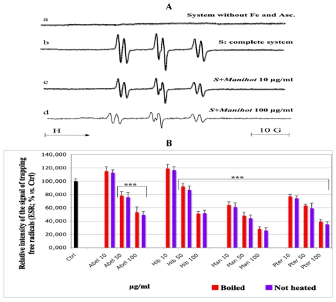
Figure 3.
Effect of the aqueous extracts of the tested plants on the formation of transient free radicals by lipid peroxidation (ESR technique). One hundred microliters of solutions of the plant extracts at the final concentrations given under the histograms were charged into 500 µL of emulsion of the linoleic acid 0.32 mM. Vitamin C 1 mM and 50 mM POBN were added. The volume was completed to 1 mL with chelexed phosphate buffer (pH 7.4) and 5 × 10⁻⁵ M Fe(II). The tubes were hermetically closed and stored at 37 °C in the dark for 2 h. Then, the content was transferred into an ESR quartz flat cell for monitoring the formation of intermediate free radicals with an ESR spectrometer. No spin trapping signal was observed at the absence of the Fe/Vit C redox system (Figure 3A, spectrum a). The addition of the Fe(II)/Vit C system initiated the lipid peroxidation and led to a signal of spin trapping with a high intensity (Figure 3A, spectrum b). This value, measured on the complete system at the absence of the plant extracts, was set as control (Figure 3B, Ctrl, black histogram, 100% signal intensity). Except Abel and Hib at 10 µg/mL which enhanced the signal, the addition of the extract solutions at increasing concentrations decreased the intensity of the spin trapping signal (Figure 3A, spectrum c, d and Figure 3B). Samples vs. Ctrl: ** p < 0.01; ***p < 0.001. The same extract at the same concentration, not heated vs. boiled sample: no significance. Abel = Abelmoschus, Hib = Hibiscus, Man = Manihot and Pter = Pteridium.

