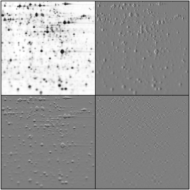
Figure 2.
A single iteration of a decimated 2-D wavelet transform with a 6-tap Daubechies wavelet on a 2-D gel region. The image is decomposed into low frequency structure (top-left), horizontal high frequency details (top-right), vertical details (bottom-left), and details from both diagonals (bottom-right). For the detail components, black represents negative values and white positive values. The diagonal detail component is scaled up by 100, which illustrates the wavelet transform’s relative insensitivity to these orientations.

