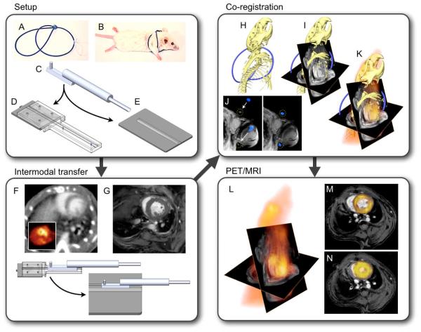
Figure 1. Experimental set up.
(A,B) Fiducial vest. (C) Multimodality imaging bed connects to gurneys on PET/CT (D) and MRI (E) to allow for a no-touch transfer of the mouse between modalities (F,G), ensuring that no major position changes hamper data fusion. (H) Three-dimensional CT data show the skeleton and a part of the fiducial marker in blue. (I) CT and MRI matrices are matched, and the angles used during cardiac MRI acquisition are applied. Then, fiducials visible on CT and MRI are aligned (J). (K) Three-dimensional data from PET/CT/MRI. (L) CT information is phased out to yield PET/MRI. (M,N) Double angulated diastolic and systolic PET/MRI short axis views.

