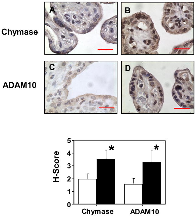
Figure 4.
Immunostaining of chymase and ADAM10 in villous tissue sections from normal and preeclamptic placentas. Both chymase and ADAM10 immnostaining were increased in tissue sections from preeclamptic placentas compared to normal placentas. Normal: A and C; PE: B and D; A and B were stained for chymase; and C and D were stained for ADAM10, bar = 20 micron, respectively. Positive staining was mainly seen in the syncytiotrophoblast layer. For the H-score graph, open column: normal; and solid column: PE, p<0.05, respectively.

