
Figure 2.
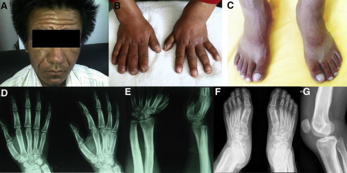
Clinical Images of the Affected Individual, Family1-P1
The images show the thickening and furrowing of facial skin (A) and the clubbing of fingernails and toenails (B and C). Hand radiographs show a loss of the normal tabulation of metacarpals and phalanges and cortical thickening of the metacarpals and the proximal and middle phalanges (D and E). A radiograph of the feet shows cortical thickening and acroosteolysis (F). A radiograph of a knee shows periosteal hyperostosis of the knee region and shows patellae sclerosis and sclerosis of both the distal femur and tibiofibula (G). All images are published with permission from the affected individual.