
Fig. 1.
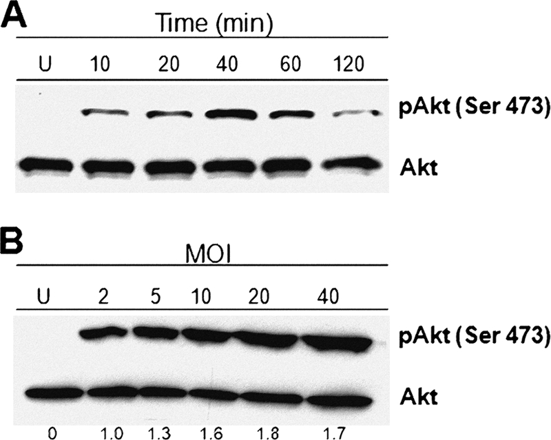
S. aureus activates the phosphorylation of Akt on Ser473 in BEC. (A) Cells were left uninfected (U) or infected with S. aureus at an MOI of 20 for 10 to 120 min. (B) Cells were left uninfected (U) or infected with S. aureus at MOIs of 2 to 40 for 40 min. After infection, the phosphorylation of Akt was analyzed by Western blotting. Detection of Akt isoforms 1 to 3 in each sample was performed to ensure equal protein loading. Blots are representative of three (A) and two (B) independent experiments. The numbers at the bottom of panel B indicate the relative band intensities obtained by densitometric analysis of each assay compared to the uninfected control.