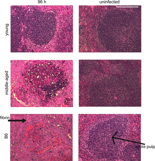
Fig. 3.
Structural integrity of spleens following Y. pestis infection. Mice were infected with ∼1,000 CFU of Y. pestis KIM5, and spleens from young B10.T(6R) mice, middle-aged B10.T(6R) mice, and B6 mice were harvested 96 h postinfection, sectioned, and stained with H&E. Images of infected (left) and uninfected (right) spleens from each individual mouse group (4 mice/group) are shown. Each image represents the findings of two independent experiments. Magnification, ×200. Bar, 0.1 mm.

