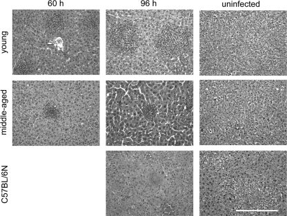
Fig. 4.
Cellular infiltrates and structural integrity of livers following Y. pestis infection. Mice were infected with ∼1,000 CFU of Y. pestis KIM5, and livers were harvested at 60 h and 96 h and were stained with H&E. At 96 h, a significant difference in the size of immune cell lesions between young and middle-aged mice is visually apparent. Necrotic lesions are found throughout the livers of B6 mice. Four mice per group were used, and each image represents the results of two independent experiments. Magnification, ×200. Bar, 0.1 mm.

