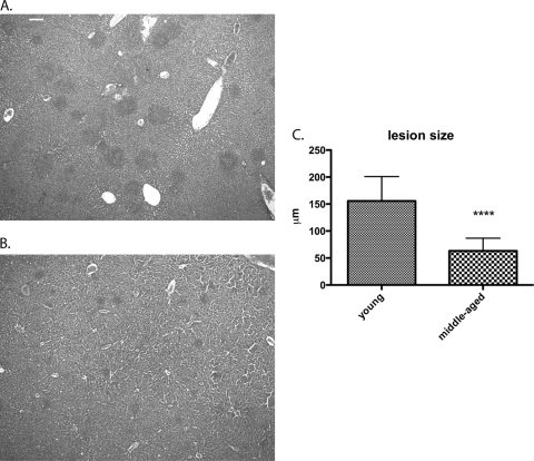
Fig. 5.
Quantification of liver immune cell lesions in young and middle-aged B10(T6R) mice. Livers from young (A) and middle-aged (7- to 9-month-old) (B) B10.T(6R) mice were harvested 96 h postinfection, sectioned, and stained with H&E. The difference in lesion size between the sections is visible. (C) Thirty lesions from each strain were measured (diameter) and compared using an unpaired two-tailed t test. Error bars represent standard deviations. Immune cell lesions were significantly larger in young B10.T(6R) mice (P < 0.0001). Magnification, ×40. Bar, 0.1 mm.

