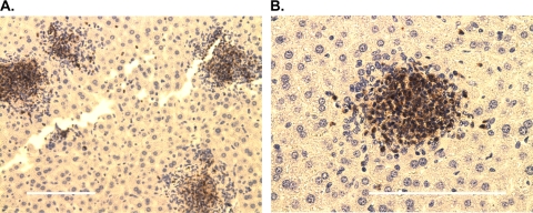
Fig. 6.
Characterization of immune cell infiltrates in the livers of young Y. pestis-infected B10.T(6R) mice. Livers were harvested at 60 h postinfection, sectioned, stained for neutrophils, and counterstained with H&E. Staining revealed tight formations of cells consisting mostly of neutrophils. Some individual neutrophils were found dispersed throughout the liver. Images represent results from two independent experiments each for four mice. Magnification, ×200 (A) and ×400 (B). Bars, 0.1 mm.

