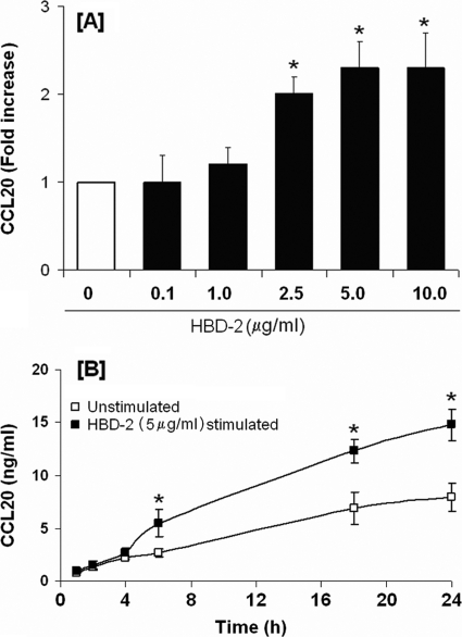
Fig. 4.
hBD-2-induced release of CCL20 by HOECs. (A) HOEC monolayers (mixed donors; N = 3) were challenged with hBD-2 (0.1 to 10 μg/ml) for 6 h; released CCL20 in cell-free supernatants was determined by ELISA, followed by calculation of the fold increase above baseline. (B) HOEC monolayers (mixed donors; N = 3) were challenged with hBD-2 (5 μg/ml) for the indicated time periods, and the amount of released CCL20 in cell-free supernatants was determined by ELISA. Results are the means ± standard deviations of three independent experiments. *, P < 0.05 with respect to control (no treatment).

