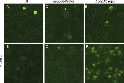
Fig. 2.
Macrophage death resulting from intracellularly induced YopJ protein. BMDMs were either left uninfected (UI) (A and B) or infected with ΔyopJ containing pVector (C and D) or pYopJ (E or F) for 20 min. After treatment with 8 μg/ml of Gm for 1 h, IPTG was included in the medium during the last 5 h of infection for half of the wells (B, D, and F). Cell death was detected by fluorescence microscopy following staining with propidium iodide (red) and annexin V conjugated with fluorescein (green). Images show the overlays of phase, and red and green signals are presented. Results shown are representative from three independent experiments.

