Fig. 8.
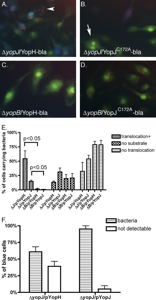
Detection of BMDMs containing translocated Yop-bla fusion proteins and intracellular Y. pseudotuberculosis. BMDMs were infected with ΔyopJ or ΔyopB strains carrying pmCherry and pYopH-bla (YopH-bla) or pYopJC172A-bla (YopJ-bla), as indicated. IPTG induction and substrate loading were carried out as described in the legend to Fig. 7. Overlaid red, green, and blue images were captured sequentially by fluorescence microscopy. (A to D) Representative images from three independent experiments are shown. (E) The percentage of cells that carried mCherry-positive (red) bacteria that were also positive for translocation (blue, translocation+) or negative for substrate loading (neither green nor blue, no substrate) or the percentage of cells in which translocation was not detectable (green, no translocation) was determined by scoring the cells in multiple images. (F) The percentage of blue cells that were positive (bacteria) or negative (not detectable) for red intracellular bacteria was determined by scoring the cells in multiple images. Results shown are the means and SEMs of three to four independent experiments. P values of less than 0.05 by the Mann-Whitney test are indicated.

