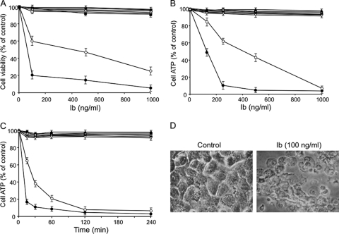
Fig. 1.
Cytotoxicity and ATP depletion induced by Ib. (A) Cells were treated with various amounts of Ib at 37°C for 4 h. Cell viability was determined via an assay with MTS, and the number of live cells is shown as a percentage of the value for untreated controls. (B) Cells were incubated with various amounts of Ib at 37°C for 4 h before ATP was measured. (C) Cells were incubated with Ib (250 ng/ml) at 37°C for the periods indicated before ATP was measured. Data are reported as percentages of the values obtained with untreated controls (means ± standard deviations for four independent experiments). Symbols (A, B, and C): ●, A431; ○, A549; ■, MDCK; □, Vero; ▵, DLD-1; ▲, CHO; ▿, HT-29; ▼, Caco-2. (D) Morphological changes of A431 cells upon treatment with Ib. A431 cells were cultured without or with Ib (250 ng/ml) at 37°C for 4 h. The cells were observed by phase-contrast microscopy. Magnification, ×150.

