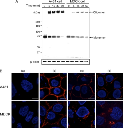
Fig. 2.
Binding and formation of oligomers by Ib in A431 cells and MDCK cells. (A) Cells (1 × 10⁶/well) were incubated with Ib (1 μg/ml) at 4°C for 1 h. The cells were rinsed, incubated at 37°C for the period indicated, and subjected to Western blot analyses of Ib and β-actin (control). A typical result from one of three experiments is shown. (B) Binding and internalization of Ib in A431 cells and MDCK cells. Cells were treated without (a) or with (b) Ib (1 μg/ml) at 4°C for 1 h. After washing, cells were incubated with medium only (c) or with medium containing Ia (d) at 37°C for 30 min. Cells were fixed, permeabilized, and stained with anti-Ib antibody and DAPI. Ib (red) and nucleus (blue) were viewed with a confocal microscope. The experiments were repeated three times, and a representative result is shown. Bar, 5 μm.

