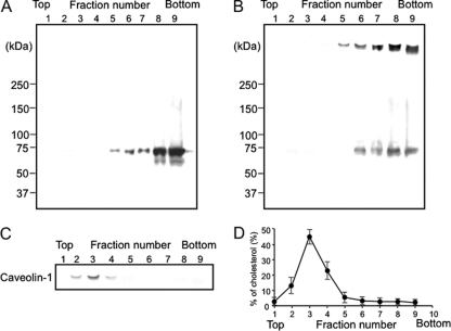
Fig. 3.
Sucrose density gradient analysis of Ib-bound A431 cells. A431 cells were incubated with Ib (1 μg/ml) in DMEM containing 10% fetal calf serum at 4°C for 1 h and then either analyzed (A) or washed, incubated at 37°C for 30 min, and then analyzed (B). Cells were solubilized in 1% Triton X-100, and detergent-insoluble fractions were floated on a step sucrose gradient. Aliquots of the nine fractions from the gradient were analyzed by Western blot analysis using anti-Ib antibody (A and B) or anti-caveolin-1 antibody (C). (D) The distribution of cholesterol in the sucrose gradient fractions was determined as described in Materials and Methods.

