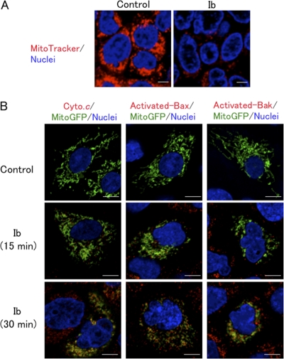
Fig. 5.
Ib induces mitochondrial dysfunction. (A) A431 cells prestained with MitoTracker red and Hoechst 33342 were incubated with Ib (250 ng/ml) at 37°C for 15 min and observed under a confocal microscope. (B) A431 cells transfected with Mito-GFP were incubated with Ib (250 ng/ml) at 37°C for the periods indicated. Cells were then fixed, permeabilized, and stained with anti-cytochrome c antibody, active-form-specific anti-Bax antibody, or active-form-specific anti-Bak antibody. Nuclei were stained with DAPI. Cells were observed by confocal microscope. The images are representative of four experiments. Bar, 5 μm.

