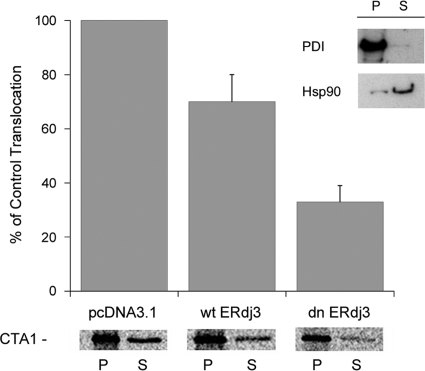
Fig. 2.
Role of ERdj3 in CTA1 translocation. CHO cells were cotransfected with plasmids encoding an ER-localized CTA1 construct and either nothing (pcDNA3.1), wtERdj3, or dnERdj3. After metabolic labeling, cell extracts were separated into organelle (pellet [P]) and cytosolic (supernatant [S]) fractions by selective permeabilization of the plasma membrane with digitonin. The distribution of CTA1 immunoprecipitated from each fraction was visualized and quantified by SDS-PAGE with PhosphorImager analysis. The averages ± standard deviations from 3 independent experiments are presented in the graph. The inset shows a Western blot control documenting the distributions of soluble ER protein PDI and cytosolic protein Hsp90 in the pellet and supernatant fractions.

