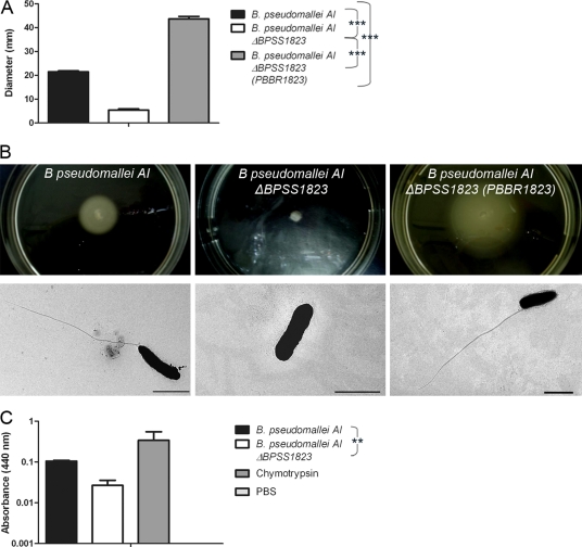
Fig. 5.
Swarming motility and protease production of B. pseudomallei AI and B. pseudomallei AI ΔBPSS1823. (A) Diameters of bacterial spread through 0.3% agar. (B) Photographs of bacterial spread through 0.3% agar and representative electron micrographs showing flagella. Scale bar, 2 μm. (C) Protease activity of bacteria using azocasein as a substrate. Values are the means from triplicate experiments ± standard errors. P values are shown for the comparison of strains.

